


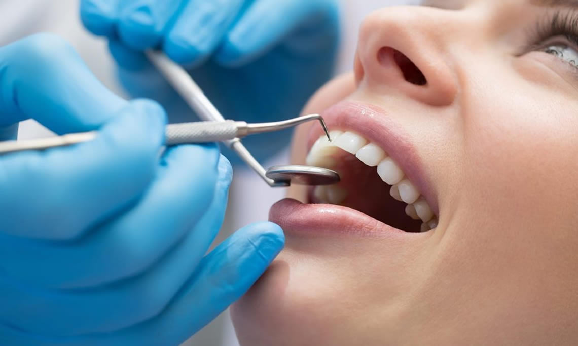
The first examination and planning are crucial for the success of a treatment. At Clinic 29, our patients receive precise diagnostics and treatment services in a professional and sterile environment.
Before treatment planning, a detailed panoramic imaging method is employed, supplemented by periapical imaging if necessary. Most importantly, the 3D DENTAL TOMOGRAPHY METHOD, which is rarely found in dental clinics, is used. Alongside radiological examination, an in-depth oral examination is conducted, and a separate unit has been established for the initial examination.
All our patients are thoroughly informed about diagnoses and treatments. Panoramic and computerized tomography radiographs are taken in our clinic to diagnose problems, plan and shape treatments. Panoramic dental X-rays are widely preferred in modern dentistry because, unlike standard X-rays, they capture the entire mouth in a single image. Additionally, panoramic X-rays can easily detect various issues, from problems in the bone structure to gum infections. You can examine the features, reasons for use, and advantages of the dental volumetric tomography device within Clinic 29 below:
Dental Volumetric Tomography (3D VT)
A dental tomography device converts data obtained from the jaw and teeth region into three-dimensional images, providing a digital representation. The acquired images facilitate the diagnosis of diseases.
Dental Volumetric Tomography (3D VT) is a newly developed tomography device designed specifically for the head and neck region. The patient can be visualized in a tomographic three-dimensional manner with minimal exposure to radiation. The detailed examination of the targeted area from the desired angle and in all directions is ensured.
When to Have Dental Tomography (3D Computerized)?
- In dental and jaw diseases,
- In dental implant applications,
- In cases of jaw-face region traumas,
- To examine in detail the relationship of embedded teeth with neighboring anatomical structures,
- For detailed diagnosis in dental and jaw pathologies,
- Before treatment planning for dentomaxillofacial surgical applications,
- For preparing surgical templates for implant placement,
- To examine anatomical structures in detail,
- To measure bone quality and density,
- To examine jawbone contours,
- To examine the temporomandibular joint,
- To examine the periapical region and canal fillings in root canal treatments,
- To examine pathological formations such as cysts and tumors,
- To examine root fractures.
Advantages of Dental Tomography (3D VT)
Planning dental implants in three dimensions before placement helps prevent future complications and allows for an adequate response to patients’ aesthetic expectations.
Three-dimensional visualization of the vertical and horizontal dimensions of the bone to be implanted provides the dentist with safer information, reducing the risk of unwanted complications in implant procedures.
Imaging is performed with up to 1/6 less radiation compared to medical tomography.
The amount of radiation given is only equivalent to 0.5 to 1.5 panoramic X-rays.
Image quality and resolution are very high.
Areas that cannot be captured with panoramic and periapical X-rays can be visualized.
Different sections can be taken from the captured image for more detailed diagnoses.
High-resolution data can be obtained for implant planning and surgical guide preparation.
Pre-treatment planning, such as soft tissue detection, bone density measurement, and measurement of mandibular canal distance, can be performed.
Postoperative controls can be conducted after treatment, prosthetics, and implants.
Detection of embedded teeth, cracks, abnormal canal numbers, and the spread of cysts can be made.

Dr. Kıvanç Türkoğlu

Dr. Pınar Türkoğlu

Dr. Başak Topuzoğlu

Dr. Lebriz Yalçın Gürhan

Dr. Sinem Kösem Mihmar

Dr. Dt. Gürcan Çetin